Figure 5.
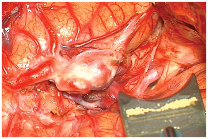
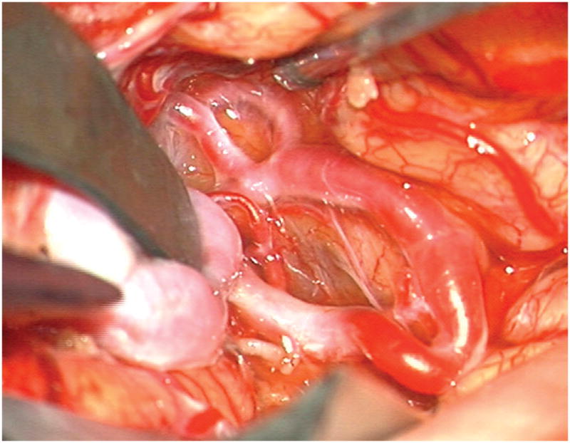
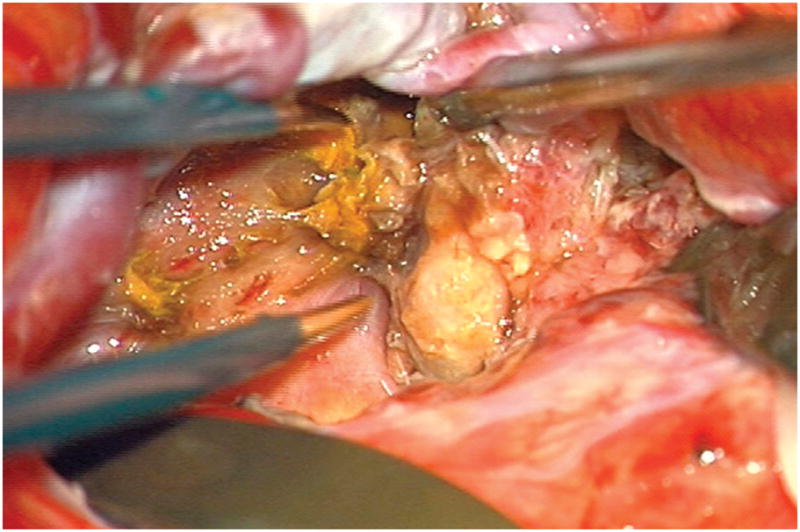
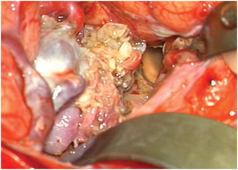
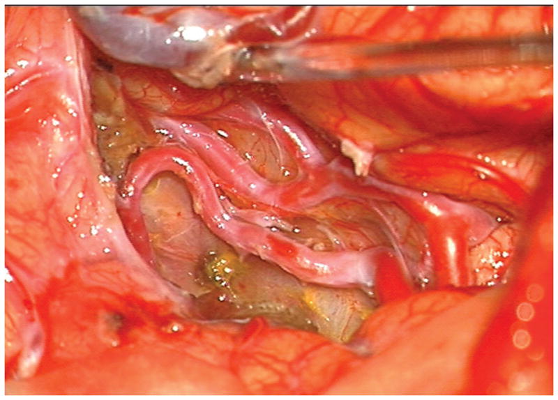
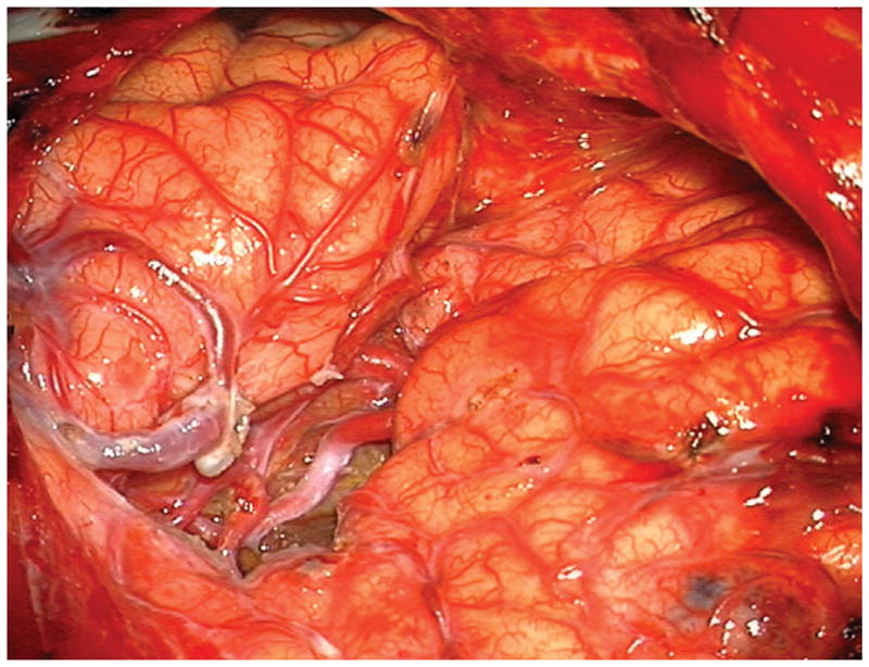
(A) Widely splitting the left proximal Sylvian fissure exposed the draining venous varix, MCA branches, supraclinoid ICA, and optic nerve (Patient 5). (B) Retraction of the varix exposed the MCA bifurcation and its branches, and the inferior margin of the AVM was visualized deep to the M2 MCA segments in the limen insulae. (C) A gliotic plane of dissection around the anterior border to the nidus resulted from previous hemorrhage and radiosurgery. (D) The medial border of the nidus connected to the lateral ventricle. (E) En passage and uninvolved arteries in the operculum were carefully preserved, as seen after AVM resection. (F) The anterior transsylvian-transinsular approach preserved language cortex and the patient awoke with no new language deficits.
