Figure 6.


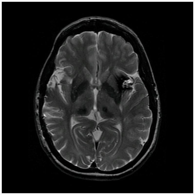
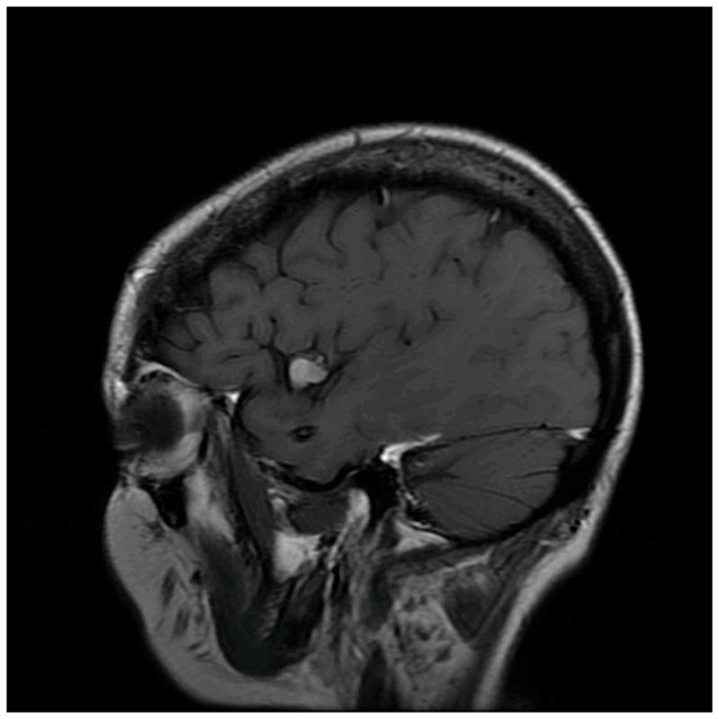
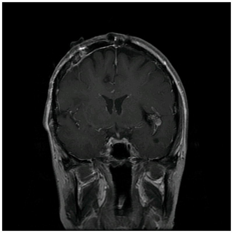
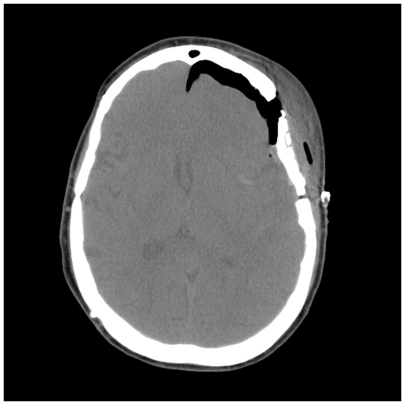
Patient 40 presented with seizures from this left insular cavernous malformation, seen on (A) axial T2-weighted, (B) sagittal T1-weighted, and (C) coronal T1-weighted MR images. The CM was resected completely through a posterior transsylvian-transinsular approach, (D) as seen on postoperative axial CT scan.
