Figure 7.
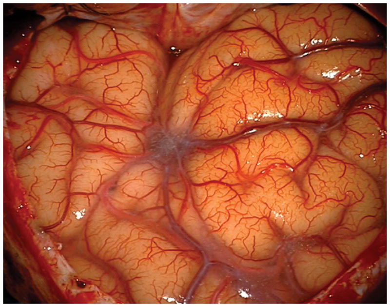
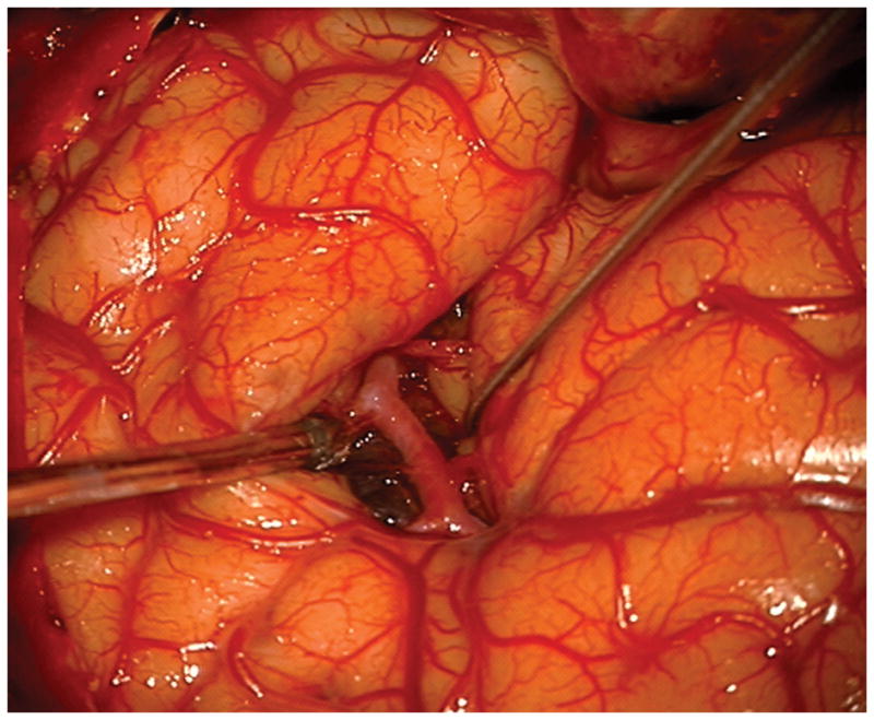
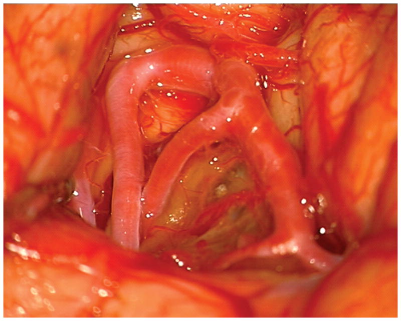
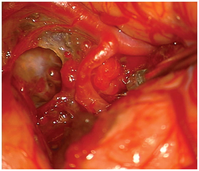
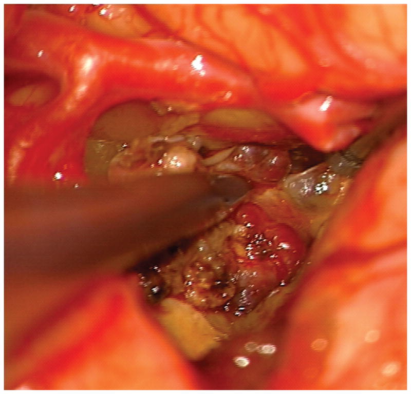
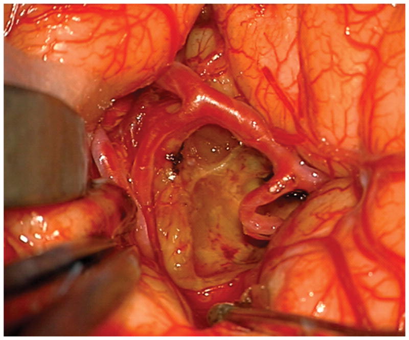
(A) The absence of overlying Sylvian veins facilitated splitting the distal Sylvian fissure, (B) which exposed the opercular segments of MCA (Patient 40). (C) The cavernous malformation came to the surface of the long gyrus and was apparent under the arterial branches. (D) Evacuation of intracapsular hematoma decompressed the lesion and facilitated the dissection in the gliotic plane. (E) The posterior transsylvian-transinsular approach preserved perisylvian cortex and overlying MCA branches.
