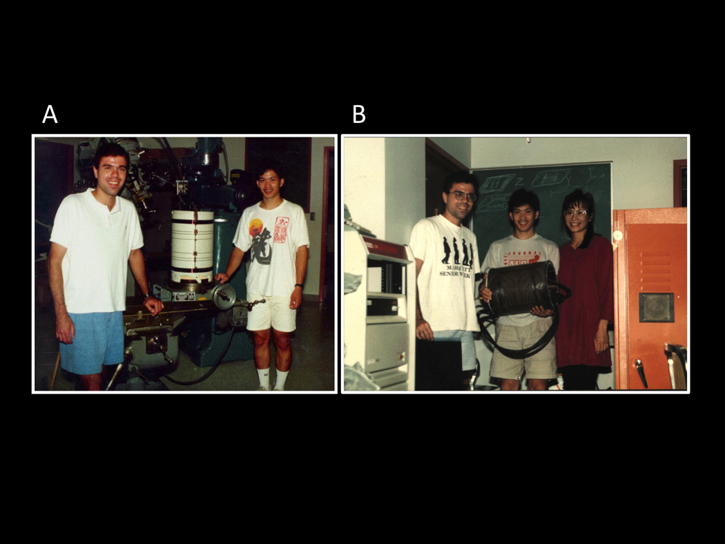
Figure 1.

These were taken during the first week of August, 1991. They show: A. Eric Wong and myself during the early stages of the construction of the three-axis balanced torque human head gradient coil that was used for the first collection of fMRI data at MCW. Here, the coil has the first layer of wires (z-axis) applied. B. Here, Eric, his wife Denise, and I show the coil after collecting the first image (an apple) with it. It was interfaced to a GE 1.5T Signa scanner. This picture was taken after we’ve been working almost continuously for about 36 hours to complete it.
