
Figure 5.
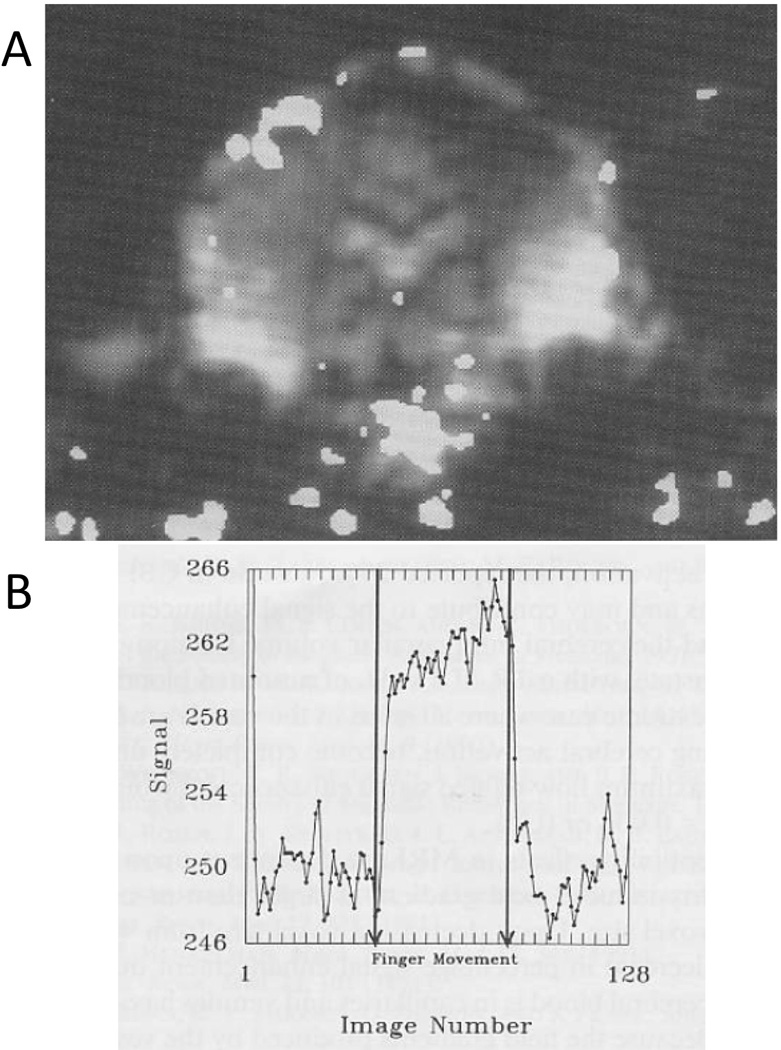
These are the results from the GE prototype z-axis head gradient coil. During this experiment (Alan Song was the volunteer), I had to pull a string attached to his hand to get him to tap his fingers. A. This is a functional map that was “thresholded” or “threshheld” (these really should become words since they are used so much in fMRI) and superimposed on an EPI anatomic image from the time series. Because the gradient coil was z-axis, we had to collect either sagittal or coronal EPI slices. Note the large pulsation artifact at the base of the brain. B. The signal to noise of the GE body RF coil was a better than that of the first generation RF coil inside the gradient coil. The time series signal from a region of interest in the motor cortex looks less contaminated by noise. (Reproduced with permission from Wiley)