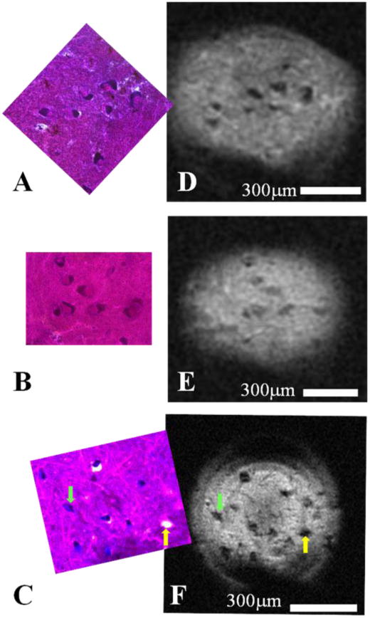
Figure 1. Human cell bodies of α-motor neurons detected using magnetic resonance microscopy and confirmed through correlative histology.

Diffusion-weighted (b = 2000s/mm2) MRM (n = 3) taken in the ventral horn of human spinal cord enlargements using a 500μm micro surface-coil and their corresponding correlative histology. MR images (DEF) show perikarya of human α-motor neurons which appear dark in contrast to the surrounding tissue of the ventral horn's gray matter. Nissl-stained histology images (ABC) contain the same cell bodies whose cytoplasmic compartments have been darkly stained due to the presence of Nissl bodies. The nuclear compartment is visible in a fraction of cells present in the histology and can be identified by its relative lack of Nissl staining and the presence of a nucleolus; however, subcellular compartments have not been visualized in the corresponding MRM. Interestingly, as is seen in panels C and F, cell bodies (green arrow) can share morphological and contrast characteristics with microscopic tissue tears (yellow arrow) at the resolutions employed.
