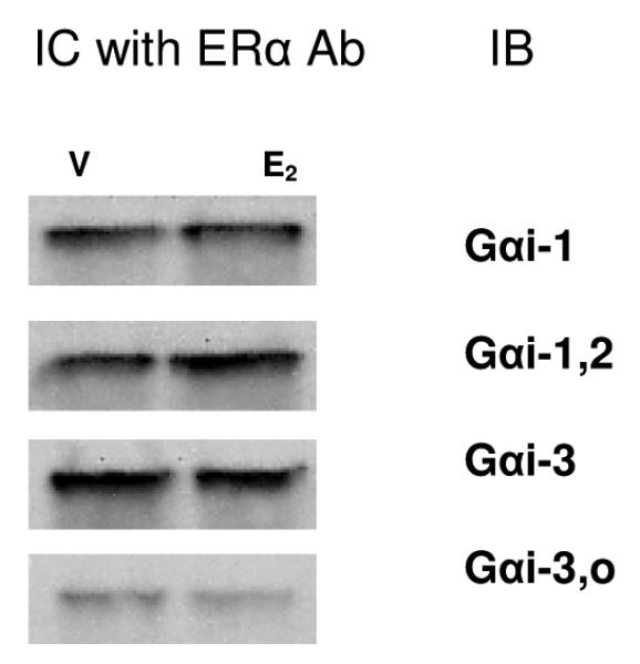
Fig. 4. ERα and Gαi proteins interact in whole-cell lysates.

Lysates from cells treated with either vehicle (V) or 10nM E2 (E2) for 10 min were bound to specific Ab to ERα (MC-20 recognizing the C-terminus) and captured on Protein G agarose beads. Immuno-captured (IC) proteins were subsequently eluted, electrophoresed, blotted, and probed with four different subtype-selective Gαi Abs (IB), as indicated. All captured G proteins were ~48 MW, and no changes were noted due to E2 treatment. A representative immunoblot from 3 experiments is shown.
