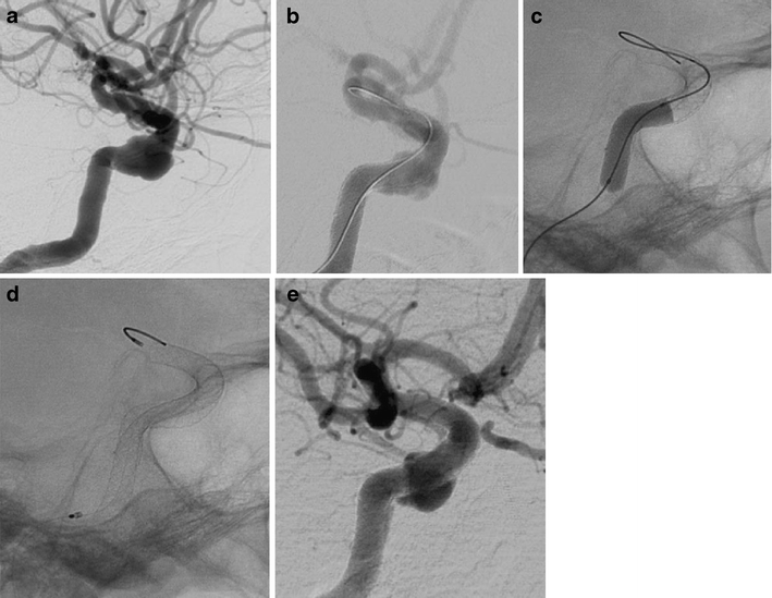
Fig. 2.

Cavernous aneurysm of the right ICA (a). Poor wall apposition after deployment of the second PED [2 × 4.5/20] (b). Adaptation of the PEDs to the vessel wall using a 4 × 15-mm Ascent balloon (c) results in secondary expansion of the second device with close contact between its outer surface and the vessel wall (d). Angiographic follow-up 3 months later shows a decrease of the aneurysm size and a regular reconstruction of the parent artery (e)
