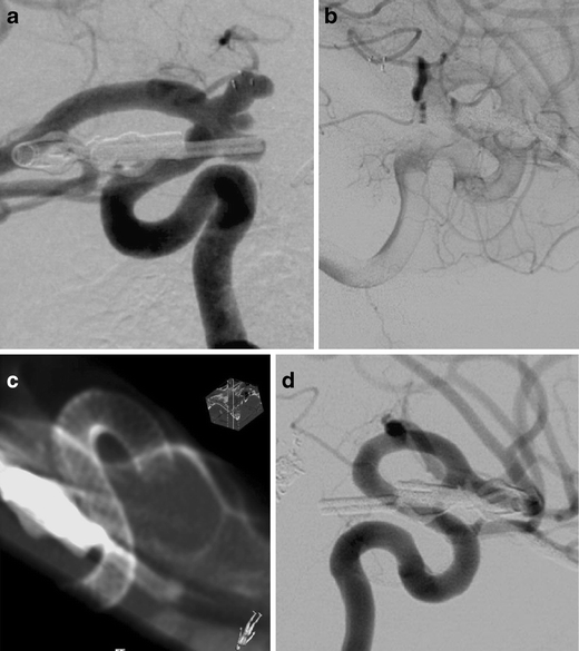
Fig. 6.

Four de novo aneurysms of the right paraclinoid and supraclinoid ICA, following microsurgical clipping of an aneurysm in the same location years ago (a). Stasis of contrast material in the aneurysms after deployment of two PEDs [2 × 2.5/20 mm] (b). DynaCT confirmed the full expansion of the PEDs (c). Obliteration of the four aneurysms with reconstruction of the lumen of the right ICA was confirmed at the first follow-up DSA after 4 months (d)
