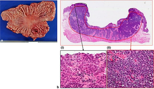
Fig. 2.
a Gross pathological examination showed that the tumor was a 3.5 × 2.5 cm type 2 lesion in the middle of the posterior wall of the stomach body. b At microscopic examination the tumor comprised two elements at the histological level (hematoxylin-eosin staining, ×2): (i) A moderately differentiated adenocarcinoma in the superficial portion of the mucous membrane layer (×40). (ii) NEC-like cells with dark, round nuclei and scant cytoplasm, presenting a solid and trabecular pattern, in the submucosal and muscularis propria layers (×40).

