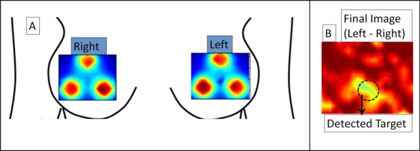
Figure 6.
Simultaneous bilateral imaging using Gen-2 hand-held imager showing (a) 2D surface contour plot of intensity distribution on left and right breast (b) final subtracted image of left-right breast, in a normal breast with a 0.45 cc absorption target embedded under left breast flap at 6’o clock position. Final image in (b) differentiates target distinctly from background artifacts, with max absorption (in yellow) at target site.

