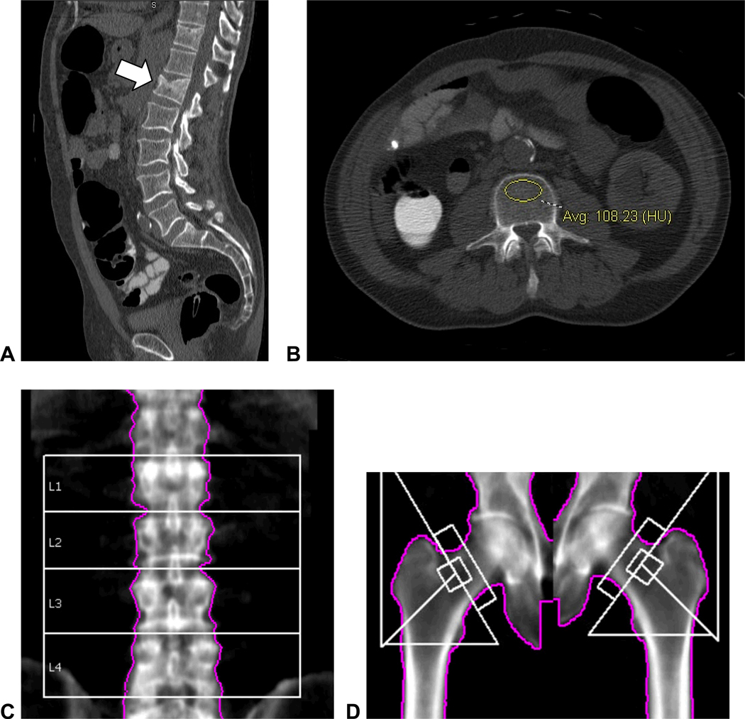
Figure 7. Unsuspected moderate L1 compression deformity in 60-year-old man with abnormally low vertebral attenuation but without osteoporosis by DXA.
Sagittal reconstruction of supine CTC series (A) shows a moderate compression fracture at the L1 level. Vertebral attenuation was abnormally low at all levels (B, 108 HU at L3) except for L1 (173 HU, not shown) due to the compression deformity itself. The reported T-scores at central DXA were 0.2 for L1–L4 (C) and −1.1 for the left femoral neck (D). Increased density at L1 level at DXA in C was attributed to degenerative changes.

