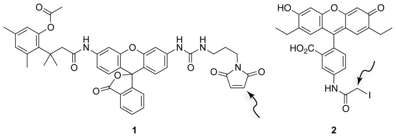
Figure 4.
Structures of fluorogenic label 1 for monitoring endocytosis (Lavis et al., 2006) and fluorescent probe 2 for monitoring protein–ligand interactions (Lavis et al., 2007). The arrows indicate electrophilic carbons that can form thioether linkages with cysteine residues.

