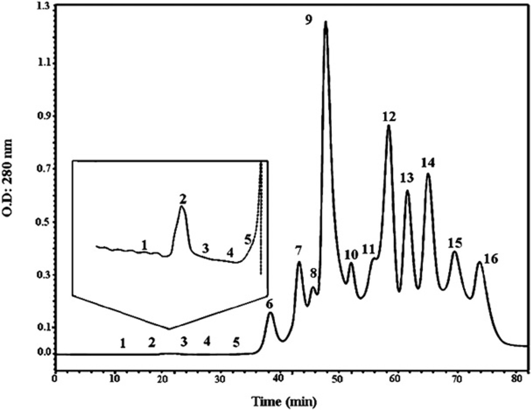
Fig. 2.
Molecular exclusion chromatographic profile of M. t. tener venom. Crude venom (5 mg) was reconstituted in 100 µl of 50 mM ammonium acetate buffer, pH 6.9 and fractionated through a Superdex-200 column (10 × 300 mm) previously equilibrated with the same buffer. The venom was run for 80 min at 0.4 ml/min, and the proteins were detected at 280 nm. The insert shows the fractions F1–F5, which showed the fractions with a high anti ADP-inducer-platelet aggregation and anti-plasmin activities.

