
Figure 1. Wnt16 is required for specification of HSCs.
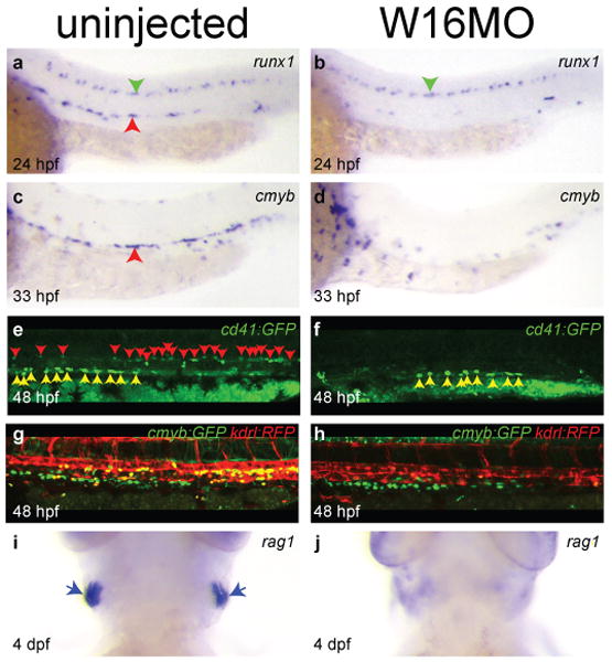
Expression of the HSC markers runx1 (a-b) and cmyb (c-d). Fluorescently labelled HSCs in cd41:gfp (e-f) and cmyb:GFP;kdrl:RFP transgenics (g-h). Expression of the lymphocyte marker rag1 (i-j). Embryos are either uninjected (left column) or injected with 5 ng W16MO (right column). Red arrowheads identify the aorta region (a, c) or individual HSCs (e). Green arrowheads identify unaffected runx1+ neurons (a-b). Yellow arrowheads identify unaffected GFP+ multiciliate cells of the pronephros (e-f). Yellow cells are HSCs (g). Blue arrows identify thymic T cells (i). a-h dorsal up, anterior left. i-j ventral views, anterior up.