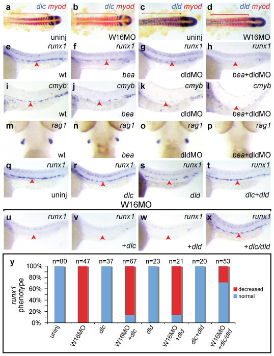
Figure 3. Wnt16 acts upstream of Notch ligands Dlc and Dld.

Expression of somitic dlc (a-b) and dld (c-d) but not myod (red) is decreased at 17 hpf in W16MO-injected embryos. Red bars indicate somites. Expression of the HSC markers runx1 at 24 hpf (e-h) and cmyb at 36 hpf (i-l) is reduced in dlc mutant (bea) embryos (f, j) or dldMO-injected embryos (g, k), and eliminated in the combined animals (h, l). The lymphocyte marker rag1 at 4.5 dpf is present in wild-type (m), bea (n), and dldMO-injected animals (o), but eliminated in bea embryos injected with dldMO (p). Combined injection of dlc/dld rescues HSCs in wnt16 morphants (q-x). Runx1 at 24 hpf. One group of embryos was uninjected, or injected with the indicated Notch ligand mRNAs alone (q-t). A second group of W16MO-injected embryos was co-injected with Notch ligand mRNAs (u-x). Percentages of embryos displaying the depicted phenotypes (y). a-d flat mounts, anterior left. e-l, q-x close up lateral views, anterior left, dorsal up. m-p ventral head views, anterior up. Red arrowheads indicate the aorta region.
