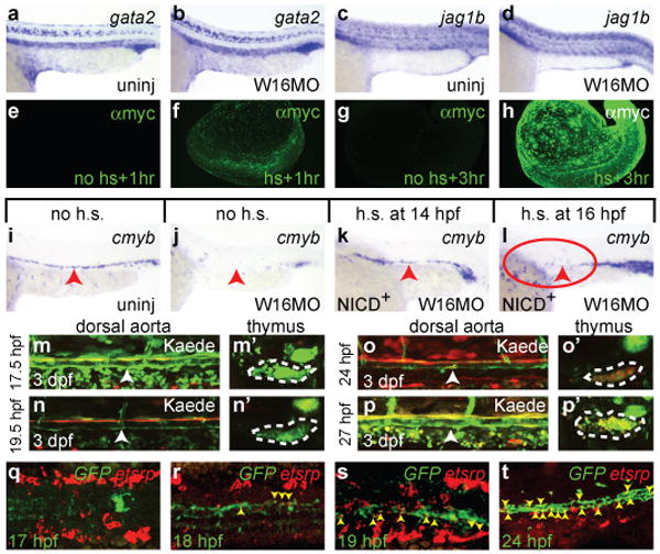
Figure 4. Non-cell-autonomous requirement for Notch in HSC specification.

Expression of the Notch target gata2 (a-b) and the Notch ligand jag1b (c-d) in uninjected (a, c) or W16MO-injected (b, d) embryos at 22 hpf. Whole mount immunofluorescence visualization of the myc-tagged NICD at 1 (e-f) and 3 (g-h) hours following either no induction (e, g) or heat-shock induction (f, h). Cmyb expression at 36 hpf in transgenic animals carrying a heat-shock inducible dominant activator of Notch signalling (NICD) in uninjected (i), W16MO-injected (j), W16MO-injected and heat-shock induced at 14 hpf (10-ss; k) or 16 hpf (14-ss; l). Red arrowheads indicate the dorsal aorta region. Red circle where HSCs should normally be expressed (h). Green-to-red photoconvertible Kaede Notch reporter animals were entirely photoconverted at the times indicated at left of each panel pair and imaged at 3 dpf (m-p′). Confocal images of the dorsal aorta (white arrowheads; m-p) and thymus (dashed white outline; m′-p′) reveal photoconverted cells only in the thymi of fish converted at 24 and 27 hpf (o′, p′). Max-projection confocal images of the trunk region of embryos processed by double fluorescence in situ for a Notch reporter GFP transgene (green) and the haematopoietic mesoderm marker etsrp (red) at the times indicated (q-t). Yellow arrowheads indicate double-positive cells (r-t). a-d, i-l close up lateral views of the trunk region, anterior left, dorsal up. e-h, whole embryo views. m-p close up lateral views of the dorsal aorta. m′-p′ single thymic lobes. q-s, dorsal views, anterior left. t, close up lateral aorta view, anterior left, dorsal up.
