
Figure 1.
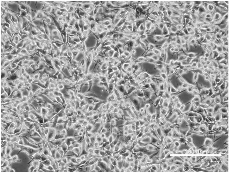
Morphology of the HS-RMS-2 cell line in culture, photographed with a phase contrast microscope. The white bar indicates 200 μm.
Official websites use .gov
A
.gov website belongs to an official
government organization in the United States.
Secure .gov websites use HTTPS
A lock (
) or https:// means you've safely
connected to the .gov website. Share sensitive
information only on official, secure websites.
Morphology of the HS-RMS-2 cell line in culture, photographed with a phase contrast microscope. The white bar indicates 200 μm.