Figure 1.
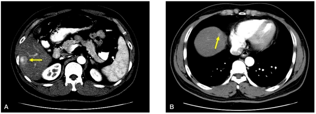
CT scans performed at the initial diagnosis of HCC. (A) The hepatic mass was a 1.7 cm-sized mass with arterial enhancement (arrow) mass in hepatic segment VI, with a patent portal vein and no metastatic lesion evident on CT scans. (B) A tiny subcapsular arterial enhancement (arrow) without early washout was noted in segment IV.

