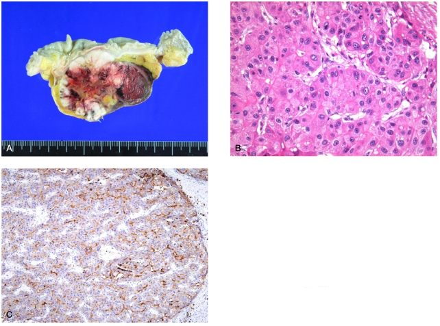
Figure 3.
Histopathology of metastatic HCC. (A) Gross specimens of sigmoid colon metastasis showing a well-defined subserosal mass. (B) The tumor cells had eosinophilic granular cytoplasms and large nuclei containing prominent nucleoli, resembling HCC (hematoxylin-eosin stain; ×400), and (C) they were positive for polyclonal carcinoembryonic antigen (pCEA) (×100).

