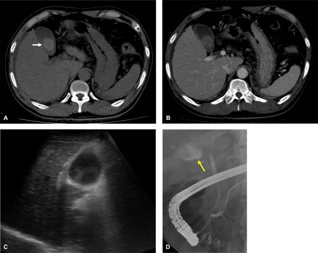
Figure 2.
A nonenhanced mass-like lesion (arrow), considered as hematoma, was seen in gallbladder after RFA. The hematoma was measured as 65.8 HU (Hounsfield units) in precontrast scan (A) and as 66.0 HU in portal-phase scan (B). Abdominal ultrasonography revealed a heterogeneous echogenic mass in the gallbladder (C). Endoscopic retrograde cholangiopancreatography showed a gallbladder with a localized filling defect (arrow) (D).

