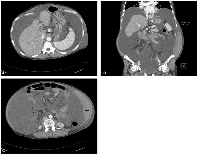
Figure 1.
(A) Abdomen computed tomography (CT) imaging after paracentesis showed severe shrinkage and irregular nodularities of the liver contour and massive ascites without evidence of hemoperitoneum. (B) Coronal abdomen CT imaging showed a large hematoma of the left abdominal wall from the margin of the rib to the inguinal area. (C) Transverse spiral abdomen CT imaging revealed a large hematoma of the left abdominal wall and focal extravasation of the contrast medium (arrow), suggesting bleeding of an abdominal wall arterial branch.

