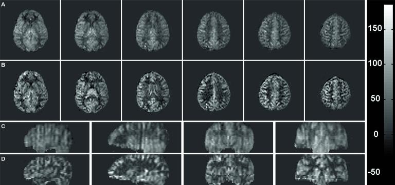
Figure 5.
Correction for blurring shows improved localization of the perfusion signal. Six transverse slices and reformatted sagittal and coronal slices obtained with (A, C) constant flip angle and (B, D) modulated flip angle are shown. Scale on right is in ml/100g/min and window/level is the same for all images.

