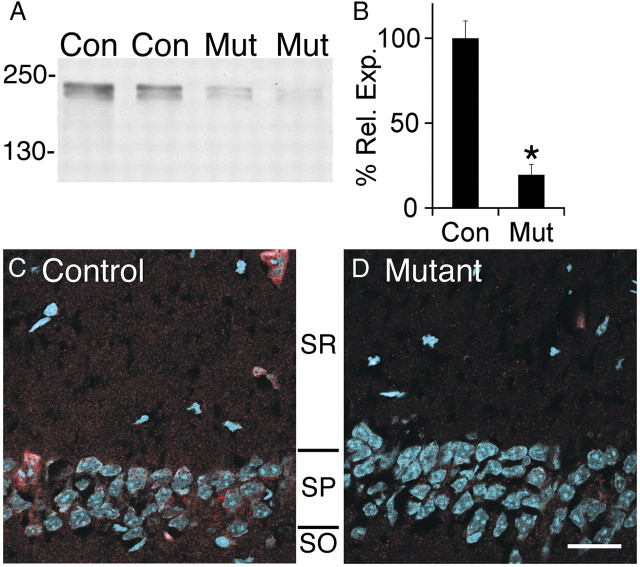
Figure 2.
Afadin expression is reduced in the hippocampus after Nex–cre-mediated deletion. A, Western blots for afadin in control (Con) and mutant (Mut) hippocampal lysates demonstrate a significant loss of both the long and short afadin isoforms. Molecular weights are listed in kilodaltons. B, Quantification of afadin protein blots in mutant (Mut) versus control (Con). Individual blots were normalized to total protein. Error bars depict SEM; *p < 0.0001; n = 6. C, D, Decreased afadin expression in mutant (D) versus control (C) demonstrated by immunocytochemical visualization of l/s-afadin (red) and nuclei (blue) in CA1–SR and CA1–SP. Scale bar, 25 μm.

