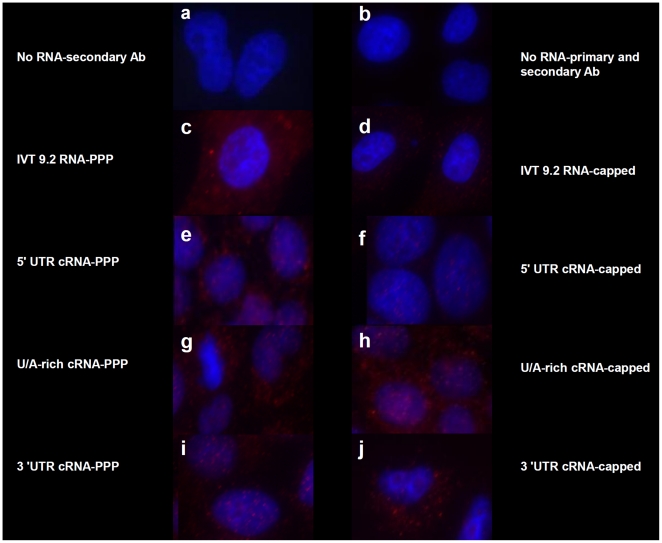
Figure 6. 5′ PPP-independent activation of RIG-I.
A549 cells were transfected with 1 µg of the indicated IVT RNAs as in Figure 2. After 24 hrs, the cells were fixed with 4% paraformaldehyde and permeabilized with a 0.2% saponin/0.1% BSA/PBS buffer. Cells were blocked using CAS overnight and probed with a conformational dependent rabbit RIG-I polyclonal primary antibody that detects the RNA-bound form of RIG-I in the cytosol and followed by staining with AlexaFluor goat anti-rabbit 549 (stains red) and Hoechst 33342 (stains nucleus blue). Cells were visualized using a Zeiss fluorescent microscope with an axiocam HRM apotome attachment using AxioVision software.

