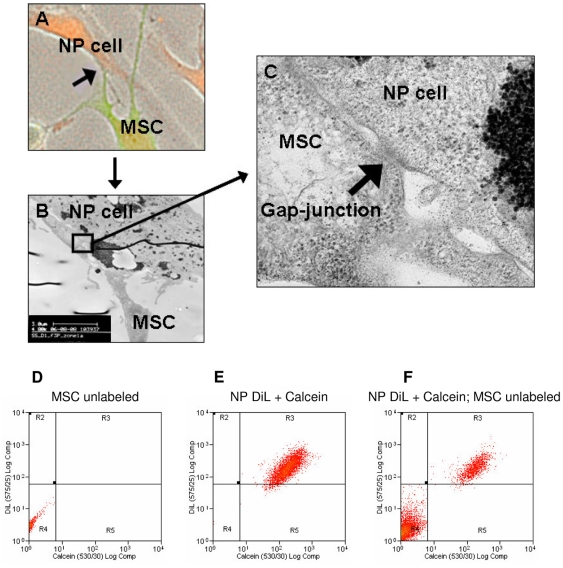
Figure 2. Formation of gap-junctions.
Example images of gap-junctions: A) Fluorescence microscopy to illustrate a potential site of cell-to-cell contact (arrow) between MSCs (green) and NP cells (red). B) TEM of the site of cellular contact between the MSC and NP cell identified in panel A. C) Enlargement of area (blocked in panel B) depicting cell-to-cell contact between MSC and NP cell revealing a typical gap-junctional structure (arrow). Example flow cytometry dot plots to identify gap-junctional dependent dye transfer between MSCs and NP cells: D) Unlabeled MSCs. E) DiL and calcein labeled NP cells. F) Direct co-culture of unlabeled MSCs and double labeled NP cells after 24 hours. No calcein only labeled cells were detectable. Abbreviations: MSC: mesenchymal stem cell; NP: nucleus pulposus.

