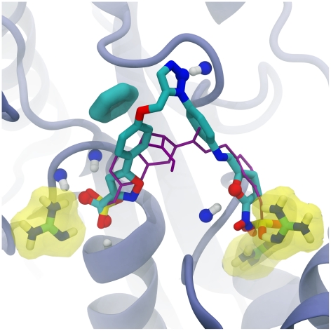
Figure 2. The top-scoring predicted PTP1B ligand (in licorice representation), docked into the receptor active site.
Protein residues that participate in electrostatic interactions are highlighted in yellow. Atoms that participate in receptor-ligand hydrogen bonds are shown in ball-and-stick representation. The aromatic ring of the receptor tyrosine residue that participates in π-π stacking and T-stacking interactions with the ligand is shown in thick licorice representation. The crystallographic pose of a known inhibitor is shown in purple, with key sulfonate moieties shown colored by element in licorice representation. Portions of the protein have been removed to facilitate visualization.

