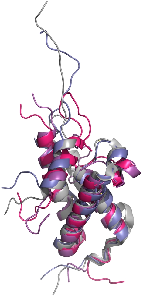
Figure 6. The loop behavior in Glu626Ala and of Leu628Ala mutants.
Alignment of the average KIX structure from the simulations of Glu626Ala mutant of KIX∶MLL complex (pink), Leu628Ala mutant of KIX∶MLL complex (purple) and KIX∶MLL structure from the NMR structure (grey) (PDB ID: 2AGH [11]) onto the average structure obtained by the KIX∶MLL wild type simulation (blue) shown using PyMOL [44].

