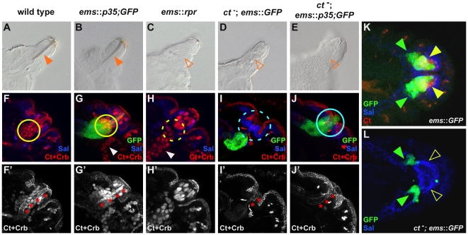
Figure 2. Cut-dependent repression of apoptosis is required for cell survival and differentiation.
(A–E) Cuticle preparations of the different genotypes with focus on the PS of 1st instar Drosophila larvae. Closed, orange arrowheads in (A) and (B) mark the filzkörper, whereas open, orange arrowheads in (C, D and E) indicate the absence of this structure in the respective genotypes. (F–L) Labeling of the different parts of the PS primordium of stage 15 embryos in the different genetic backgrounds using the filzkörper marker Ct (red, nuclear), the stigmatophore marker Sal (blue) and the apical membrane marker Crb (red). In (G, I, J, K and L) GFP expression (green) driven by the ems-GAL4 driver is shown in the different genetic backgrounds. Red asterisks in (F′–J′) mark the invaginated cells of the future filzkörper. Closed, yellow circles in (F) and (G) mark Ct-positive, invaginated filzkörper precursor cells, dashed yellow circle in (H) indicates the absence of these cells. Dashed, light blue circle in (I) highlights the absence of GFP-positive cells, whereas closed, light blue circle in (J) mark the presence of these cells. Note that some cells expressing GFP under the control of the ems-GAL4 driver invaginate deeper than the Ct expressing cells, thus they are still present in ctdb7 mutant embryos, indicated by closed, green arrowheads in (K) and (L). Closed, yellow arrowhead in (K) marks Ct and GFP-positive cells in ems::GFP embryos. Open, yellow arrowhead in (L) highlights the absence of these cells in ct mutant embryos. In (A) to (J′) lateral views, in (K) and (L) dorsal views of embryos are shown.

