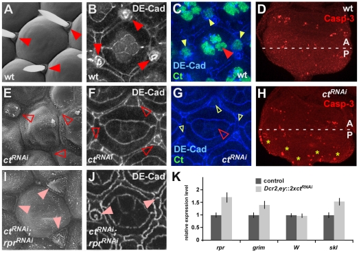
Figure 3. General function of Ct in apoptosis repression and induction of differentiation.
(A, E, I) Scanning electron micrographs of individual ommatidia of adult Drosophila fly eyes with indicated genotypes are shown. The closed, red arrowheads in (A) mark interommatidial bristles, the open, red arrowheads in (E) mark the absence of these structures. The closed, light red arrowheads in (I) indicate the presence of tissue that would normally develop into interommatidial bristles. (B, F, J) Projections of consecutive confocal sections of one ommatidium of 50 h pupal retinas labeled with DE-Cadherin. Interommatidial bristles are marked by red, closed arrowheads in (B). Open arrowheads in (F) mark absence of DE-Cad, light-red arrowheads in (J) mark reduced DE-Cadherin levels in shaft cells of interommatidial bristles. (C, G) Projections of consecutive confocal sections of one ommatidium of 50 h pupal retinas of GMR::lacZ control (C) and GMR::ctRNAi flies (G). (D, H) Expression of the apoptosis marker Caspase-3 (Casp-3) in 3rd instar eye-antennal discs of control Dcr2; ey::lacZ (D) and Dcr2; ey::2xctRNAi (H) animals. Yellow asterisks in (H) mark Casp-3 positive cells in Dcr2; ey::2xctRNAi eye imaginal discs. (K) Relative mRNA expression levels of rpr, grim, Wrinkled (W) and sickle (skl) in 3rd instar eye-antennal discs of control Dcr2; ey::lacZ and Dcr2; ey::2xctRNAi animals.

