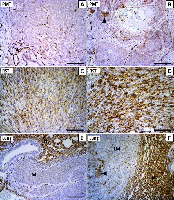
Figure 2.
Cav-1 immunohistochemistry in PMT tissue (A, B), RST tissue (C, D) and two different lung metastases (E, F). The arrows in (A) indicate Cav-1 positive cells lining blood vessels in normal mammary tissue while T marks the mammary tumor. The arrowhead in (B) highlights Cav-1 positive tumor cells within a PMT sample. In panels (E, F) LM identifies the lung metastasis while NL identifies normal lung tissue. Scale bars, 100 μm

