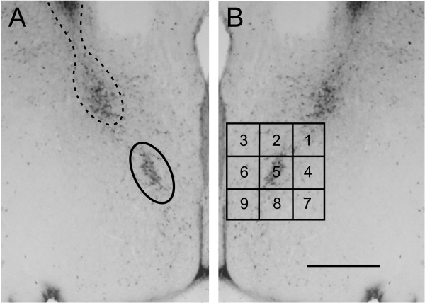
Figure 1.
Ellipse and grid contours designed to quantify calbindin-immunoreactive (ir) cell number in and surrounding the calbindin-ir sexually dimorphic nucleus in the preoptic area (CALB-SDN). The photographs in (A) and (B) are mirror-image duplications of a single image from a male mouse. (A) For CALB-SDN counts, an ellipsoid contour (solid line) was placed as indicated. A second calbindin-ir cell cluster was found dorsolateral to the CALB-SDN. These cells were determined to be the ventral extension of the bed nucleus of the stria terminalis (BNSTp) (dotted line) and were not included in CALB-SDN counts. (B) For counts of cells within and surrounding the CALB-SDN, a nine-block grid was placed as shown and counts were made within each block of the grid. Scale bar = 400 μm.

