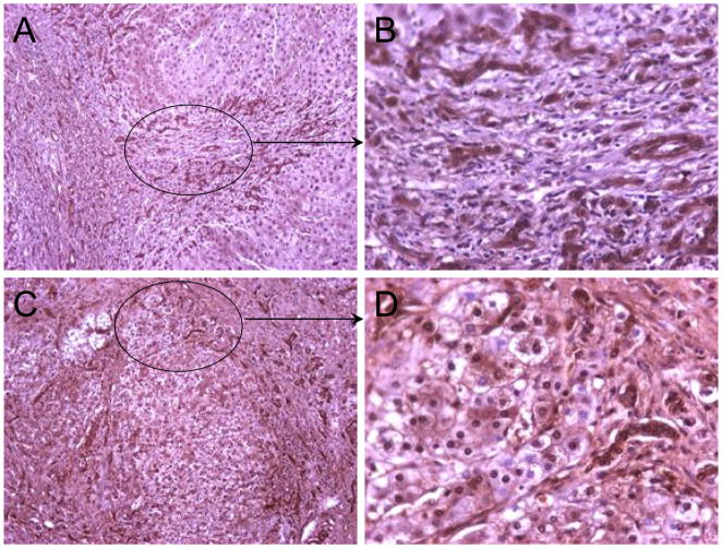
Fig. 3. Localization of DcR3 expression in dysplastic nodule.
The upper panels (A & B) show a case of pure cirrhosis with no atypia. Inside the cirrhotic nodules, the cells were practically negative for DcR3. The lower panels (C & D) present a case of cirrhosis with marked atypia which could be regarded as early cancer; cells inside the nodule were mostly DcR3 positive. Both DcR3 positive (dark brown) and negative (light blue) nuclei can be observed in panel D. Original magnification: A, C, x100; B, D, x400 (DAB, haematoxylin counterstain).

