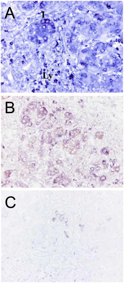
Fig. 4. In situ hybridization of DcR3 transcripts in CHC with cirrhosis.

In the areas of active inflammation and architectural remodelling (panels A & B), there is intense staining in infiltrating lymphocytes (Ly), as well as in bile ductular and intermediate cells (I). Images in B and C (inset) were processed simultaneously to show the background level staining in CHC cirrhosis with sense-probe (negative control). Brown granules seen in panels B and C are hemosiderin. Original magnification: A, B, and C, x400.
