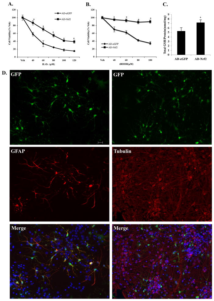
Figure 3. Overexpression of Nrf2 selectively in astrocytes protected neighboring neurons against toxins in DJ-1 KO primary cortical neuronal cultures.
Cells were transduced with 50 MOI (multiplicity of infection) adenovirus-eGFP-Nrf2 (AD-Nrf2) or adenovirus-eGFP (AD-eGFP). Forty-eight hr later cultures were treated with either A. H2O2 or B. tBOOH. Mean±SEM, n=5. *Significantly different from corresponding AD-eGFP value (two way ANOVA, p<0.05). C. Total intracellular glutathione levels in primary cortical cells were measured after a 48hr infection of 50 MOI AD-Nrf2 and AD-eGFP. Mean±SEM, n=7. *AD-Nrf2 vs AD-eGFP (two-tailed t test, p<0.05). D. Infection of primary cortical cultures with 50 MOI of adenovirus showed adenovirus-infected cells colocalized with GFAP-positive astrocytes and, rarely with β(III)-tubulin-positive neurons (100 MOI) (scale bar 20μm). Blue: Hoechst 33258 staining.

