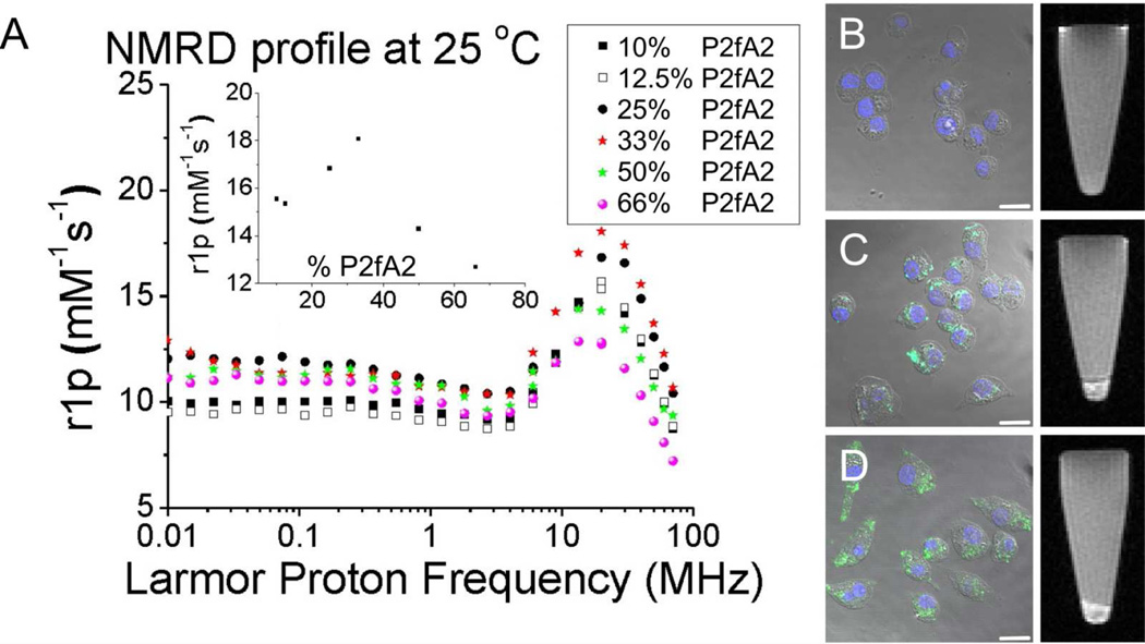
Figure 2.
(A) NMRD-profiles of the different structures. The inset displays the relaxivity as function of %P2fA2 at 20 MHz. Confocal microscopy (left) and MRI of loosely packed cell pellets (right) of macrophage cells that were (B) left untreated, incubated with 33% (C) and (D) 50% P2fA2 nanoparticles. Scale bar: 20 µm.

