Figure 6.
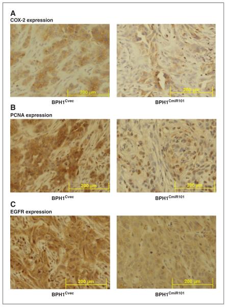
Expression of COX-2, PCNA, and EGFR in BPH1CmiR101 and BPH1Cvec tumor xenografts. The expression levels of COX-2 (A), PCNA (B), and EGFR (C) in the BPH1CmiR101 tumor xenografts were compared with the corresponding levels in BPH1Cvec by immunohistochemical staining method. As described under Materials and Methods, the tumor xenografts were removed at the end of the experiment, fixed in formalin, and then stained with specific monoclonal antibody. Magnification × 400.

