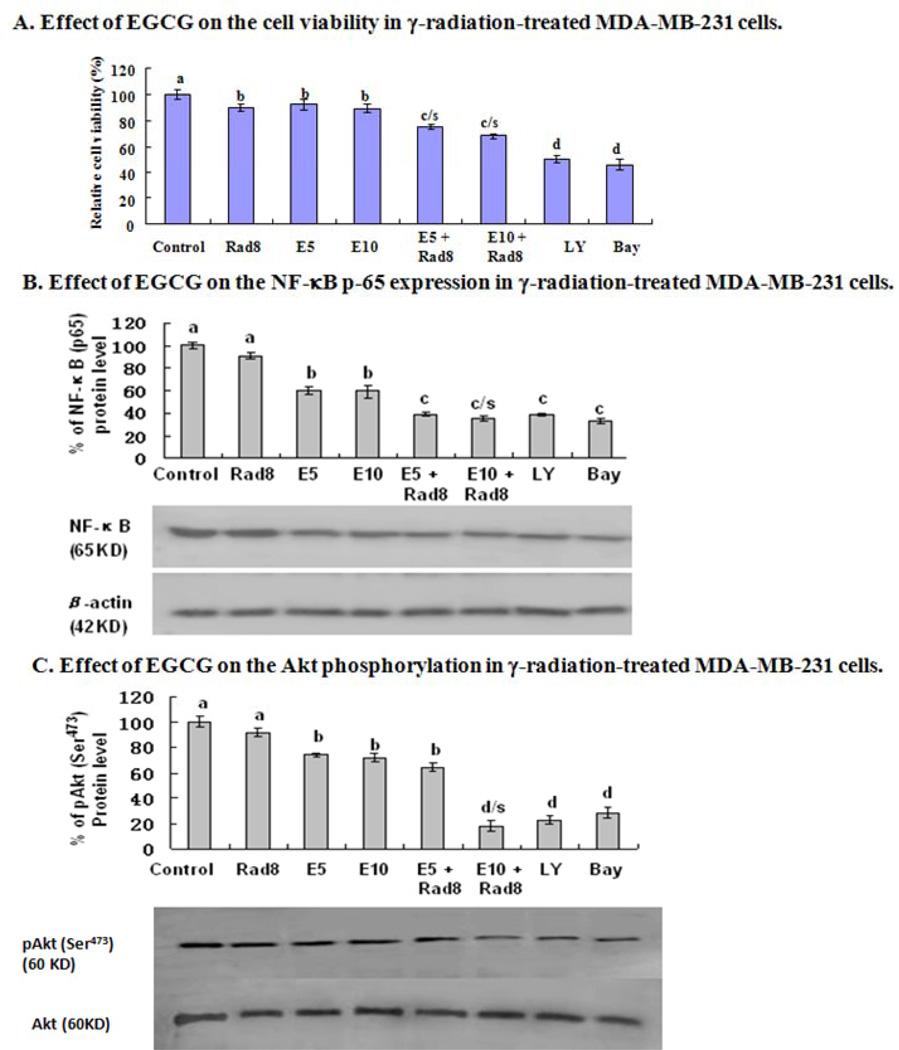
Fig. (7).
In vitro effects of EGCG on cell viability and protein level of NF-κB p65 as well as phosphorylation (Ser473) of Akt in γ-radiation-treated human breast cancer cells. MDA-MB-231 cells were treated with γ-radiation at a dose of 8 Gy (Rad8) in the absence or presence of EGCG at concentrations of 5 µM (E5) or 10 µM (E10). After the treated cells were cultured for 36 h, the relative cell viability was determined using a trypan blue dye exclusion assay (A); the equal amounts of protein from total cell lysates were separated by SDS-PAGE, and Western blot analysis of NF-κ B p-65 (B) and p-Akt (C) were done. Bay 11–7082 (BAY), the inhibitor of NF-κB, and Ly294002 (LY), the inhibitor of PI3K/Akt were used as a positive control, respectively. The density of the band (normalized to β-actin for NF-κB or to Akt for p-Akt) shown as mean ± SD is relative to that of the control (designated as 100%). Statistical analysis was carried out using the two-way ANOVA followed by Bonferroni post hoc test to establish whether significant differences existed between the groups. Values with different letters (a–d) differ significantly (P < 0.05). c/s and d/s represent significantly enhanced γ-radiation effects by EGCG treatment compared with the treatment either with γ-radiation or EGCG alone (c/s, P < 0.001, two-way ANOVA; d/s, P < 0.0001, two-way ANOVA).

