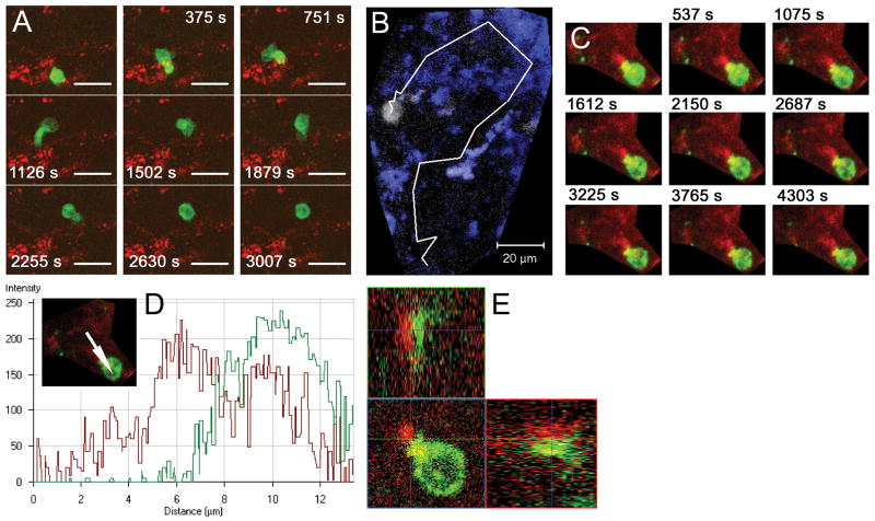
Figure 4. CD4+ T cells interacting with MHC class II+ APCs in the subarachnoid space.
Leptomeningeal explants were prepared from naive mice (A) or mice immunized with MOG35-55 harvested before onset of neurobehavioral signs of EAE (B–E) and imaged using confocal time-lapse microscopy directly ex vivo. Figures show merged z-stacks evenly spaced in time. (A) A highly motileCD4+ cell (green) migrating in a scanning fashion and forming serial brief interactions with multiple MHC class II+ APCs (red). (B) CD4+ cell (white) from mouse harvested immediately before onset of EAE migrating with higher instant velocity than cell from naive mouse (A), but retaining a scanning migration pattern and interacting with multiple MHC class II+ cells (blue). Image shows CD4+ cell trajectory outlined in white. (C) Stable interaction between CD4+ cell (green) and MHC class II+ APC (red) lasting throughout the scanning time of 70 min. (D) Pixel intensity histogram (for path indicated with arrow in insert) and (E) orthogonal 3D display of interaction in panel C showing intimate contact between the two cells. Scale bars: A–B=20 μm; C=10 μm. Time-lapse animation of cells in A–C are available in Supplementary Movies 1 and 2.

