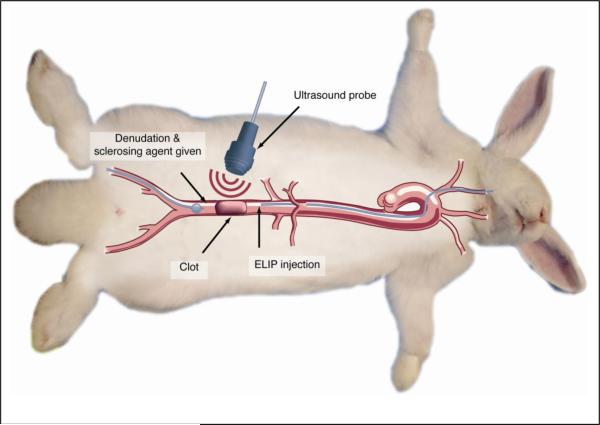
Figure 1.
Diagrammatic representation of procedures comprising the rabbit aorta thrombus model. Sequentially, these procedures include 1) denudation of the abdominal aorta, using an embolectomy catheter introduced into the right femoral artery; 2) measurement of baseline abdominal aortic flow by spectral Doppler echocardiography; 3) induction of a thrombus by administration of 5% sodium ricinoleate and thrombin; 4) measurement of post-thrombotic flow distal to the thrombus; 5) introduction of thrombolytic preparation proximal to the thrombus, through a catheter introduced into the right carotid artery; 6) in rabbits receiving ultrasound treatment, transabdominal administration of color Doppler ultrasound for 2 minutes or intermittently for 30 minutes; and 7) spectral Doppler flow measurements every 2 minutes for 30 minutes.

