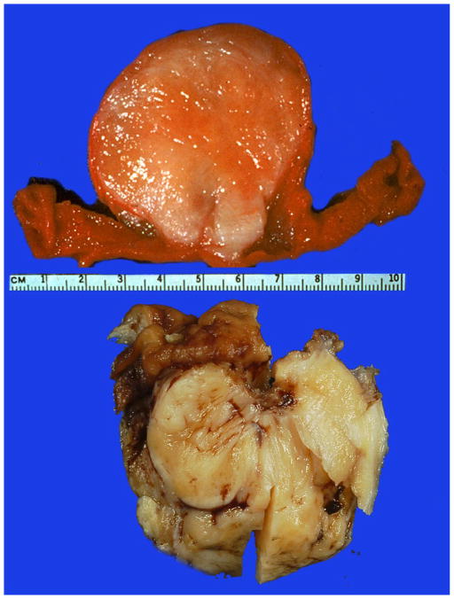
Fig 2.
Gross appearance of two gastric schwannomas. The upper panel shows and endophytic, sessile polypoid tumor with a whitish to pale tan surface of sectioning of an unfixed specimen. The lower panel contains a portion of a fixed tumor with a yellowish surface on sectioning. Mucosa is seen on top left.

