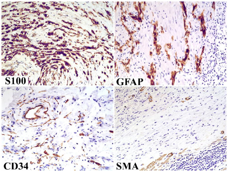
Fig 5.

Immunohistochemically typical features include positivity for S100 protein and GFAP. Both markers highlight a microtrabecular pattern. Tumor cells are negative for CD34 and SMA; the positive cells represent non-tumor elements: fibroblasts/endothelial cells and smooth muscle cells/pericytes, respectively.
