Figure 3.
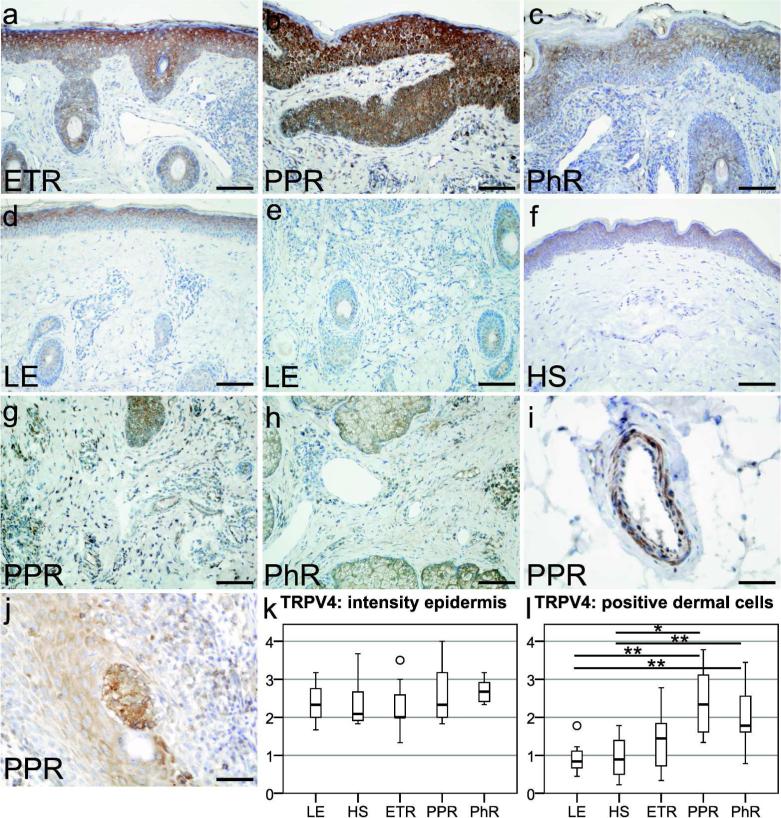
Localization of transient receptor potential vanilloid subfamily 4 (TRPV4) in erythematotelangiectatic rosacea (ETR), papulopustular rosacea (PPR), phymatous rosacea (PhR), lupus erythematosus (LE), and healthy skin (HS), followed by semiquantitative analysis of TRPV4 immunoreactivity in the epidermal or dermal compartment. (a–f) TRPV4 immunostaining was located on keratinocytes in all study groups, on dermal immune cells of rosacea, and in LE patients. Certain immune cells were strongly positive in (g) PPR and (h) PhR, whereas TRPV4-positive immune cells were only rarely found in (e) LE. Interestingly, in PPR endothelial cells, (i) smooth muscle cells and (j) Langerhans cells showed immunolabeling. (k) Semiquantitative analysis of epidermal staining revealed no significant differences. (l) Semiquantitative scoring of positive dermal cells confirmed an increased staining for TRPV4 in PPR (P<0.01) and PhR (P<0.05) as compared with HS and LE. Bar=100 µm (a–h) and 50 µm (i–j); unfilled circles represent outliers; *P<0.05, **P<0.01.

