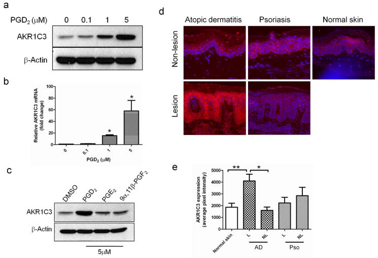
Figure 5. AKR1C3 is upregulated in AD lesions and in PHK treated with PGD2.
(a) Dose-dependent upregulation of AKR1C3 protein and (b) mRNA by PHK treated with various doses of PGD2 and harvested after 24 hours, determined by western blot (n=3) and RT-PCR (means ± SEM, n=3, *P<0.01), respectively. (c) PHK were treated with 5μM of the indicated prostaglandin for 24 hours and the regulation of AKR1C3 was assessed by western blot (n=3). (d) Immunofluorescence staining showing the relative expression of AKR1C3 in epidermis of AD (n=4) psoriasis (n=3) and normal skin (n=4). One representative case is shown, images obtained at 10x magnification. (e) ImageJ analysis of AKR1C3 staining intensity. Data is presented as the mean ±SEM of 2 separate experiments where 4 normal skin samples, 4 AD and 3 psoriasis cases were evaluated. L=lesion, NL=non-lesion, *P<0.001, **P=0.012.

