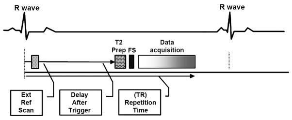
Figure 1.
Schematic of NC-MRA using ECG-triggered, T2-prepared, Fat-saturated, segmented b-SSFP with the coil sensitivity and image data acquired at two different cardiac phases (early systole and mid diastole, respectively) in the same cardiac cycle within a single BH, in order to increase the acceleration efficiency and avoid the misregistration due to varying breath hold positions.

