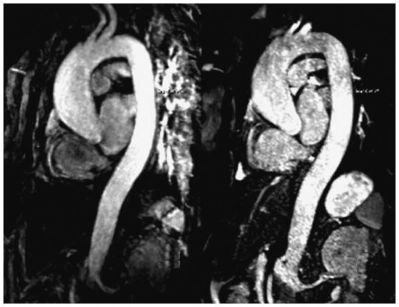
Figure 3.

MR images in a 59-year-old patient suffering from aneurysm of the aorta root obtained with NC-MRA (Right) and CE-MRA (Left) sequence. The relative merits of each technique are: CE-MRA provides superior vascular delineation due to the enhancement by contrast; whereas the single BH ECG-triggered NC-MRA is relatively free from motion artifact, so cardiac morphology is more clearly visualized, with sharper delineation of the aortic root and better assessment of coronary artery origins, due to its much shorter acquisition window (110 ms vs. 216 ms; NC-MRA vs. CE-MRA, respectively).
