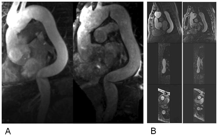
Figure 4.

A) MR images in a 79-year-old patient with tortuous thoracic aorta obtained with NC-MRA (Right) and CE-MRA (Left) sequence. The reconstruction planes clearly demonstrate sharper delineation of the aorta root and other aorta segments with high image quality for NC-MRA. B) Multiplanar reformatted images from NC-MRA (Right) and CE-MRA (Left) at the mid-descending aorta: sagittal, coronal and axial images.
