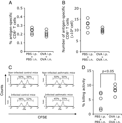
Fig. 7.
The antigen-specific cytotoxicity of CD8+ T cells following influenza virus infection. C57BL/6 mice were sensitized and challenged with OVA or PBS. Subsequently, the mice were infected with 100 pfu of influenza virus. An FCM analysis and an in vivo killing activity assay were performed on day 6 after the infection. Cells in the spleen were incubated with H-2Db Influenza NP tetramer followed by staining with both an anti-CD8 Ab and an anti-CD3 Ab and analyzed by FCM. Cells that were CD8-positive, CD3-positive, and influenza NP tetramer-positive were identified as antigen-specific CD8+ T cells (a). The number of antigen-specific CD8+ T cells was calculated by total cell counting (b). Representative FACS data of the in vivo killing assay from non-infected control or asthmatic mice and infected control or asthmatic mice are shown (c). The percentages of in vivo killing activity for asthmatic mice or control mice were calculated as indicated in the “Materials and Methods” (d). Five mice were used in each group. Similar results were obtained from two independent experiments

