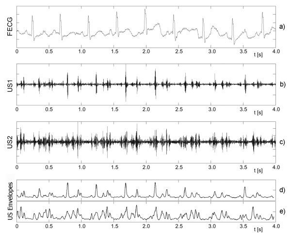
Figure 1.
Fetal heart activity signals. Four-second segments of the simultaneously acquired signals: a) direct electrocardiogram from an electrode placed on fetal head, b) and c) two Doppler ultrasound signals from two transducers placed separately but focused on the same fetus. Additionally, the envelopes of both Doppler signals are presented as d) and e). Both US signals differ significantly as for the number of cardiac cycle episodes being observed. The periodicity of Doppler signal is much easier to estimate in US1, but only the FECG signal enables explicit recognition of the timing of fetal cardiac events.

