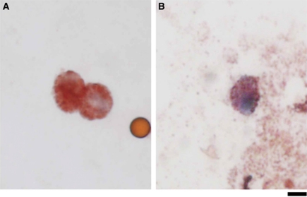
Figure 1.
Melanoma cells isolated from peripheral blood. HMW-MAA+, CD45− cells were isolated on immunomagnetic beads and stained with a mixture of MART-1 and gp100 antibodies. MART-1/gp100-positive cells stained brownish-red with aminoethylcarbazole. (A) Spiked MMG-1 cells isolated from the peripheral blood of a healthy individual. The small light-brown spheroid is an immunomagnetic bead. (B) CTC detected in a patient with melanoma. Scar bar: 5 μm.

